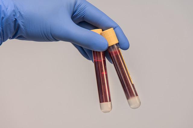
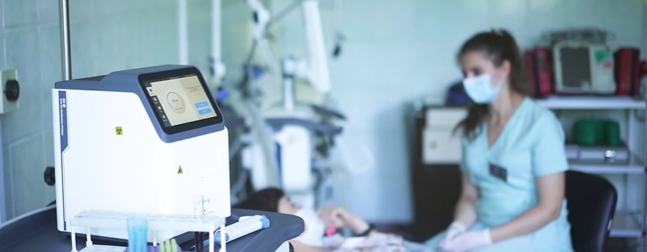
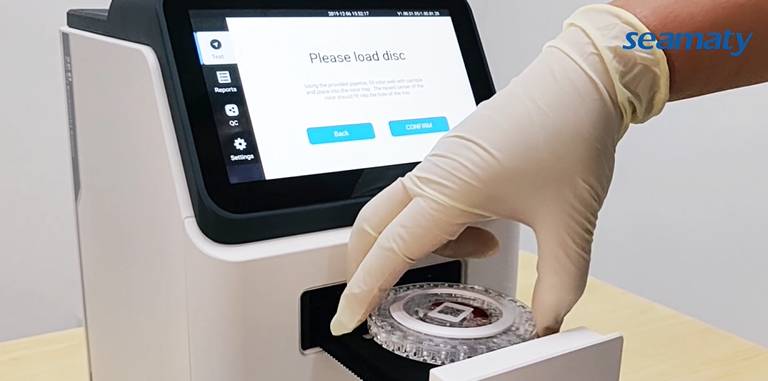
Unqualified specimens encountered in clinical testing work are mainly manifested in hemolysis, coagulation, contamination, insufficient or excessive collection volume, and incorrect identification.
The causes of substandard specimens mainly include.
-
1) the use of incorrect containers or additives.
-
2) The use of inappropriate blood collection apparatus. such as the use of undersized or oversized needle containers, the use of oversized negative pressure vacuum tubes, etc.
-
3) Incorrect specimen identification.
-
4) Irregularities in the specimen collection process causing hemolysis of the specimen.
-
5) Too little or too much blood collected.
-
6) specimen contamination. Unqualified specimens will certainly affect the test results.
01 Blood cell count and sorting count
Blood clotting or blood cell aggregation caused by poor blood collection, inadequate anticoagulation due to improper anticoagulant ratio or incomplete mixing. This can lead to low results in the corresponding cell counts. Aggregated cells may also be misclassified by the instrument as inaccurate counts of other cells.
Mixing of tissue fluid due to excessive squeezing during peripheral blood collection can lead to platelet aggregation, which can result in a low platelet count. The mixing of tissue fluid can also cause dilution of the blood.
Hemolysis caused by overly vigorous drawing and mixing techniques or improper additions. This can lead to low cell count results. During peripheral blood collection, puncture before the disinfectant is completely dry may lead to destruction of blood cells, resulting in low count results.
Blood collection at the site of inflammatory infiltration leads to local admixture of inflammatory cells, resulting in abnormal cell morphology. Adhesion and aggregation of blood cells combined lead to compromised cell sorting count results.
Blood cytology should be anticoagulated with EDTA, and the wrong anticoagulant may cause changes in blood cell morphology. This may result in inaccurate blood cell counts and classifications. For example, oxalate and heparin anticoagulation can cause low platelet count, white blood cell count and lymphocyte count results.
02 Bleeding and coagulation programs
Poor blood collection and low blood volume cause improper anticoagulant ratio or incomplete mixing, resulting in inadequate anticoagulant, leading to activation of the coagulation process and depletion of coagulation factors. This situation may cause prolongation of endogenous and exogenous clotting time and low results of some coagulation factors.
Wrong anticoagulants such as EDTA, heparin, especially heparin can cause prolongation of PT, APTT times. Poor blood sampling, prolonged pressure banding (not to exceed 30s) causing blood flow stasis or vascular damage may cause tissue factors to enter the bloodstream, which may result in lower results for some coagulation factors.
03 Erythrocyte sedimentation rate
Specimen hemolysis, poor blood collection, improper ratio of anticoagulant or incomplete mixing, etc. make clotting activated and plasma composition may change. Defects in specimen collection, such as changes in red blood cell morphology, may cause varying degrees of red blood cell aggregation, resulting in an acceleration of the hematocrit sedimentation rate. This may cause tissue factors to enter the blood, resulting in a decrease in the results of some coagulation factor measurements.
04 Biochemical items
Biochemistry mainly determines ions, enzymes and metabolic substances in the human body. The concentration of these substances varies relatively widely in different tissues and cells of the body. They are greatly affected by hemolysis. Some substances, such as enzymes and metabolites are poorly stable and easily degraded. There are also chemical methods that are inherently less specific and susceptible to interference from abnormal substances in the specimen. Therefore, the detection of biochemical items are more demanding on the specimen.
-
1) Hemolysis: When hemolysis occurs, the contents of red blood cells enter the plasma, causing changes in plasma composition. This can have a large impact on the results of some biochemical assays (such as glutamate transaminase, lactate dehydrogenase, potassium) with large differences in intra- and extracellular concentrations.
-
2) Lipid blood: lipid blood is mainly caused by the increase of celiac particles in serum, because the latter has the property of scattering light, which can produce serious interference to both colorimetric and turbidimetric methods. And this interference can not be eliminated by dual wavelength.
-
3) Jaundice: bilirubin has light absorption at 400-540 nm wavelength. The bilirubin can also be oxidized to biliverdin and biliverdin after the specimen is placed for too long or passed through oxidizing agents. These substances can also cause changes in light absorption, thus affecting the test results. For single-wavelength biochemical instruments this effect is more significant.
-
4) poor blood draw, the use of pressure pulse belt can lead to stagnation of venous blood flow, resulting in an increase in blood lactate concentration.
-
5) Blood gas analysis samples that are not drained of air or not completely isolated from air during sample collection may cause an increase in oxygen partial pressure measurements and a decrease in carbon dioxide partial pressure measurements.

05 Immunoassay Program
Immunoassays are performed by measuring a test article (antigen or antibody) by means of a labeled or unlabeled antigen-antibody reaction. Since antigen-antibody reactions are very specific, the results are relatively little affected.
However, due to the generally low level of the immunological test item being measured. If the sample is defective, especially if cross-contamination occurs, the effect on the results may be greater.
Commonly used methods for immunological assays include immunoturbidimetric methods, enzyme-labeled color development methods. such as ELISA and immunoblotting, fluorescence-labeled methods, colloidal gold-labeled spot permeation or chromatography methods. Different detection methods have different sensitivities to specimen defects.
1) Immunoturbidimetric method: the use of optical methods directly on the antigen antibody reaction produced by the precipitation detection. Therefore, it is sensitive to the color and transparency of the specimen. Severe hemolysis, lipemia and jaundice may interfere with such experiments, resulting in high results.
2) Enzyme-labeled color development technique: usually use horseradish peroxidase and other enzymes to label and catalyze the color development of the substrate. Specimens with strong reducing agent contamination (such as vitamin C infusion ipsilateral limb blood collection), will have an impact on the results, producing false negatives. In addition, because hemoglobin has peroxidase activity, severe hemolyzed specimens may also catalyze the color development of the substrate, raising the background or generating false positives.
06 Blood culture
Unqualified blood culture specimens are mainly contamination, improper ratio of blood and culture fluid, and inappropriate blood culture bottles.
1) Contamination: collection irregularities can cause contamination may occur at any stage of the blood culture operation. Numerous studies have shown that skin colonization flora is a common bacterium for blood culture contamination. It shows that incomplete skin disinfection and improper blood collection operations are the main causes of contamination.
Blood is susceptible to contamination by skin surface flora during blood collection. Mainly during peripheral venipuncture, local skin disinfection is incomplete and bacteria are cultured with the needle prick into the examined blood.
Secondly, patients with long-term indwelling vascular catheters, whose catheters are exposed to the skin outside for a long time, cause the migration of these flora. Blood cultures are also often taken from these catheters. As a result, the bacteria that migrate within the catheter are often taken out and cultured during the blood collection process.
Even with strict asepsis, it is difficult to control the contamination rate to less than 2%. Blood culture contamination will lead to unnecessary antibiotic treatment, prolong hospitalization, increase the patient's medical costs and the generation of bacterial resistance.
2) Blood and culture bottle ratio is not appropriate: adults and children blood culture blood collection standards are different, should be strictly in accordance with the manufacturer's recommended standards for collection. Too much or too little blood collection will reduce the positive rate of blood culture.
3) Inappropriate choice of blood culture bottles: The types of fully automated blood culture bottles are common bottles, neutralizing antibiotic bottles, anaerobic bottles, and children's bottles. Each type of culture bottle meets different clinical needs. Choosing inappropriate blood culture bottles will reduce the positive rate.
07 Molecular biology testing program
Failure of molecular biology specimens is commonly due to exogenous nucleic acid contamination caused by irregular blood drawing practices. Template RNA degradation and the presence of PCR inhibitors.
1) Exogenous nucleic acid contamination: due to the very high sensitivity of PCR. Theoretically, contamination of one nucleic acid molecule may cause false positive results. Therefore, it is best to use sterile, nuclease-free disposable equipment for PCR specimen collection. The collection must be strictly aseptic, and care must be taken to prevent the mixing of hair and dander from the operator or the subject. Sealed transportation and storage, specimen collection and processing should not be performed in the amplification area to prevent contamination of PCR products.
2) Target gene degradation: In general, the stability of DNA samples collected aseptically is good, and they do not affect the test when left at room temperature for 8h. However, if the sample is improperly handled, the container is not clean, left for too long or contaminated, the DNA strand may break, making the amplification of long fragments difficult. Target gene degradation has a more serious impact on RNA samples. Due to the presence of a large number of RNA enzymes in the environment, if the sample is not well preserved, transported or stored for too long, the template RNA is very easy to degrade, resulting in false negatives.
3) PCR inhibitors: Reverse transcription and PCR reactions are both enzymatic reactions. Any substance that may inhibit these reactions will affect the results of the test. Common inhibitors caused by defective sample collection include heparin, hemoglobin, lactoferrin, IgG, protease, fibrin, etc. Laboratory Medicine Network
08 Differences in analytes between peripheral blood and venipuncture samples
Although the differences in analytes between peripheral and venous blood are minimal, statistically and/or clinically significant differences exist for glucose, potassium, total protein, and calcium.
The results of hemoglobin, glucose, and potassium tests were higher in peripheral blood than in venous blood. In contrast, the results of sodium, chloride, calcium, bilirubin, and total protein tests were lower than those of venous blood. Oxygen partial pressure and oxygen saturation were lower in peripheral blood than in arterial blood. Reference ranges should be established when testing these items using peripheral blood, and the type of specimen should be indicated on the test report.
With substandard specimens, the testing is ineffective labor, even if the laboratory uses the best methods and techniques. The test results can delay the timely diagnosis and proper treatment of patients.
Therefore, the correct collection of specimens is an important and indispensable part of quality assurance in the pre-analytical phase of clinical testing, and is also the basis for ensuring accurate, reliable and valid clinical test results.


