Technical principles of Hematology Analyzer tests

release time:2022-07-20 15:54:02

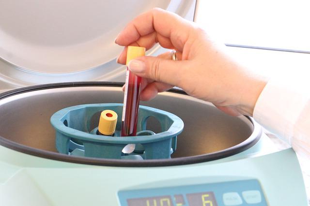
In a conventional hematology meter, red blood cells (RBC) and platelets (PLT) share a single measurement channel. The determination of hemoglobin content (HGB) is the same in any type and grade of hematology analyzer. White blood cell counting and classification has its own dedicated channel. The technical methods and principles used for each test item on the hematology analyzer are briefly described.

Technical principles of Hematology Analyzer tests.jpg

1. Hemoglobin Content Measurement

Hemoglobin content is measured by adding a hemolytic agent to diluted blood, which causes the red blood cells to release hemoglobin. The latter combines with the hemolytic agent to form a hemoglobin derivative, which enters the hemoglobin test system.

The absorbance changes in proportion to the Hb content of the liquid at a specific wavelength (usually 530-550nm). The instrument can then display the Hb concentration. Different series of hemolytic agents are formulated for different hematology analyzers. The hemoglobin derivatives they form are also different. But most of them have a maximum absorption spectrum close to 540nm.

In recent years many high-grade hematology analyzers have adopted laser scattering method for the analysis of individual blood red blood cell hemoglobin. This is to minimize the effect of high WBC, celiac blood, high bilirubin, etc. on the colorimetric HBG.

2. Hematocrit and platelet assay

The detection of red blood cells is an important part of the hematology analyzer. In the past, red blood cells were mainly counted by the impedance method in terms of number and volume. The signals of different sizes were then sorted out and a histogram of the red blood cell volume distribution was printed.

However, nowadays, a combination of optical and electrical impedance processing is also used to perform three-dimensional spatial analysis (3D) of red blood cell volumes in order to obtain more accurate results. For example, Bayer's ADVIA 120 detects red blood cells by light scattering. This hematology analyzer measures one red blood cell simultaneously with two measurement systems: low-angle forward light scattering and high-angle scattering. The volume and total number of individual red blood cells are measured based on the magnitude of the low-angle light conversion energy. The individual hemoglobin concentration is obtained from the high-angle light scattering, and the MCV (mean red blood cell volume), MCH (mean hemoglobin content) and MCHC (mean hemoglobin concentration) measurements can be accurately obtained, and the histogram of red blood cell scattering, individual red blood cell volume and Hb content in red blood cells, and the parameters of RWD (red blood cell volume distribution width) and HDW (red blood cell hemoglobin distribution width) can be plotted. distribution width) and other parameters.

Due to the obvious difference between platelet and red blood cell volumes, it is easy to distinguish the photoelectric signals measured at the same time with a limited threshold. Therefore, to date, a common analysis system has been used for platelet and erythrocyte testing in whole blood analysis. However, there is often a crossover between platelet and red blood cell measurement signals. For example, the pulse signal of a large platelet may be mistaken for a red blood cell and counted. The pulse signal of small red blood cells may enter the platelet channel. This can result in experimental errors. Various manufacturers of hematology analyzers use various advanced technologies to reduce the interference of platelet counts. We describe each of these techniques below:

Sweep Flow

Since platelets and red blood cells are counted in the same counting cell, the cell volume is large, and the platelets can be counted in the same pool. The cells are large in size and form a large pulse when they pass through the central counting induction zone. If there is reflux, a small pulse is formed at the same time due to the eddy current re-entering the edge of the sensing zone, so that the electrode may sense a small pulse equivalent to the size of platelets, resulting in a false increase in platelet count.

The sweep flow technique is to have a steady stream of fluid pass behind the red blood cell counting aperture while the red blood cell and platelet counts are being performed. This allows the red blood cells to be washed away immediately afterwards to prevent them from returning to the sensing area to be counted as platelets.

Hematology Analyzer History

The conventional method of routine hematological examination is manual testing with the aid of a microscope. After staining the blood for red blood cells, white blood cell count and blood smear, the microscope performs manual leukocyte sorting with the naked eye. It took at least 20 minutes for each specimen to come out of the test, which was not only a small number of test items, but also time-consuming and laborious, and the accuracy and reliability were affected to some extent, making it difficult to perform quality control.

Since the introduction of the Automated Heamatology Analyzer in the 1950s, automated blood cell analysis has evolved from a single electrical impedance technique to a fusion of multiple techniques. These include physics, chemistry, immunology, flow cytometry, information processing techniques, volumetric conduction light scattering (VCS), and multi-angle polarized light scattering (MAPSS). This blood technology has led to more accurate and reliable results for the analysis of various blood cells.

Automated Hematology Analyzer

Automated blood cell analysis has evolved from a single semi-automatic unclassified to 3/5-part differential to an automated blood cell analysis pipeline. The processes of complete blood count (CBC), reticulocyte (Ret) count, peripheral blood pushing and staining are fully automated.

Hematology Analyzer Clinical Applications

The testing parameters of automated blood cell analysis have developed from a single blood cell count result to provide more than twenty parameters for clinical diagnosis, differential diagnosis, treatment and prognosis monitoring.

The precision and accuracy of CBC test results have been significantly improved. Large and medium-sized hospitals have multiple hematology analyzers in their laboratory departments. Some large or teaching hospitals also have automatic blood analysis assembly line. At the same time, hospitals at all levels have basically established quality control procedures for automatic blood analysis, which has greatly improved the efficiency of whole blood cell counting.
Prev Return Next

Related Reading

Hematology Analyzer History | Principle -CBC Machine-Seamaty

2022-07-20

Hematology Analyzer, also known as Blood Cell Analyzer or Blood Cell Counter, is mainly used for testing blood specimens. This instrument can qualitatively and quantitatively analyze the blood components and provide relevant information.

How to Choose a Suitable Biochemistry Analyzer?

2022-06-07

In medicine, biochemistry analyzers are considered one of the essential tools. They are used primarily for diagnosing and monitoring diseases, but they can also be used for other purposes such as drug testing and food analysis.

Clinical significance of biochemical indicators of liver damage in cats

2021-12-17

The liver, located at the diaphragm, is one of the most important organs in the abdominal cavity. It plays a vital role in maintaining the normal functioning and metabolism of the body, such as 1. aiding digestion (metabolism of fats) 2. synthesis of proteins and hormones.