Compared with
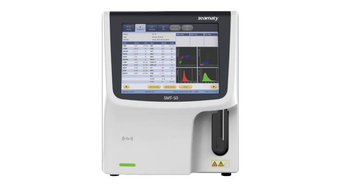
3-Part Auto Hematology Analyzer, 5-Part Auto Hematology Analyzer can better meet the clinical needs because of the increase in the routine detection of eosinophils and basophils. Therefore, many hospital laboratory departments and laboratories have chosen the 5-Part Auto Hematology Analyzer.
1. Electrical impedance, high frequency conductivity and laser scattering combined detection method
It is to do Kurt's VCS technology.
V: stands for volume, which is the electrical impedance principle invented by Kurt. The resistive microporous technique is applied to count blood cells and determine their volume.
C: stands for conductivity, which refers to a beam of electromagnetic waves or radio frequency. The conductivity of a beam of electromagnetic waves or radiofrequency, which penetrates the cell, depends on the size and internal structure of the cell. The corrected conductivity is called capacity, which no longer reflects the cell size. Instead, it reflects the internal structure of the cell. This reflects the internal structure of the cell, such as the size of the nucleus, the nucleoplasmic ratio and the density.
S: stands for scatter, which refers to the intensity of scattered light measured at different angles. The scattering of light varies between cells due to different surface structures and particle sizes, thus distinguishing between different types of cells. Kurt has designed the instrument to measure the same leukocyte by all three methods simultaneously on a single channel, eliminating the need to design several channels or add staining reagents to some hematology analyzers.
Principle of 5-Part Auto Hematology Analyzer
Based on the principle of fluid dynamics. The analyzer applies the sheath flow technique to allow the remaining leukocytes and reticulocytes after hemolysis to pass through the detector individually. It is subjected to simultaneous detection by all three VCS techniques. Finally, the leukocyte sorting process is completed.
2. Light scattering and cytochemical staining combined detection method
It uses laser scattering and peroxidase staining techniques for cell sorting. The principle of light scattering is used to count blood cells. There are differences in the scattered light at different angles due to different cell surface structures. This helps in the differentiation of leukocytes.
Leukocyte chemical staining means that a substrate of peroxidase (MPO) and a color-presenting agent are added to the dilution solution. Leukocytes are classified according to their reaction to MP0. Neutrophils are strongly positive for MP0. Eosinophils are more positive. Monocytes are weakly positive. Basophils are also negative. Therefore, combining the characteristics of light scattering and cytochemical staining, a two-dimensional or three-dimensional image similar to flow cytometry can be obtained on the computer monitor one by one with different color scatter plots. And the classification results can be typed. Since basophilic cells do not contain this enzyme, they cannot be detected.
Therefore, some blood analysis instruments are specially equipped with basophil detection system. It uses a time difference method for its measurement channel. It shares a common channel with the measurement system of RBC/PLT. Among laser technologies there are currently semiconductor lasers and gas lasers. The semiconductor laser is weak in power, but is sufficient as a cell sorting itself. Gas laser power is strong both compare semiconductor laser has the advantages of long life (7 to 10 years) small size, fast start, low power consumption, low voltage regulation requirements, more economical, etc.
3. Combined electrical impedance and RF conductivity detection method
This method uses four detection systems separately to detect different types of cells.
①Lymphocyte, monocyte and neutrophil detection system
In the cell suspension Huai add hemolytic agent to make red blood cells lysis. Leukocytes remain intact. The cell pulp and nuclear morphology approximate the physiological state. As these cells pass through the assay system, the leukocytes are tested by a combination of the electrical resistance method (measuring cell volume) and the radiofrequency conductivity method (detecting nuclei and particle density). The test results will classify the cells into three groups: lymphocytes, mononuclear fine and neutrophils.
②Two detection systems for eosinophils and basophils
A special hemolytic agent is added to the cell suspension. All cells except eosinophils and basophils are lysed or atrophied. The eosinophils or basophils that remain intact are then counted.
③Naive cell detection system
Amino acid sulfide is added to the cell suspension. Due to different occupancy and more amino acids in naive cells than in mature cells. And the naïve cells are resistant to the hemolytic agent. When the hemolytic agent is added, the mature cells are lysed. It retains only the naïve cells that may be present for counting.
4. Multi-angle laser polarized light scattering detection method
Hematology analyzers using this technique use a sheath flow solution to dilute the specimen blood. The internal structure of the diluted leukocytes approximates their natural state. Only the basophils have a slight change in cell structure due to their hygroscopic properties. The hemoglobin within the erythrocytes is separated from the cells by the high osmotic pressure. In contrast, water from the sheath flow enters the erythrocyte, leaving the cell membrane structure still intact. It has the same refractive index as the sheath flow and does not affect the detection of leukocytes.
This hematology analyzer instrument detects the scattered light from cells passing through the laser beam from four angles simultaneously. This is because cell size, refractive index, nuclear shape, nucleoplasmic ratio, and particle properties can all affect the amount of scattered light measured at different angles.
These are the 4 methods and principles of the 5-Part Auto Hematology Analyzer.


