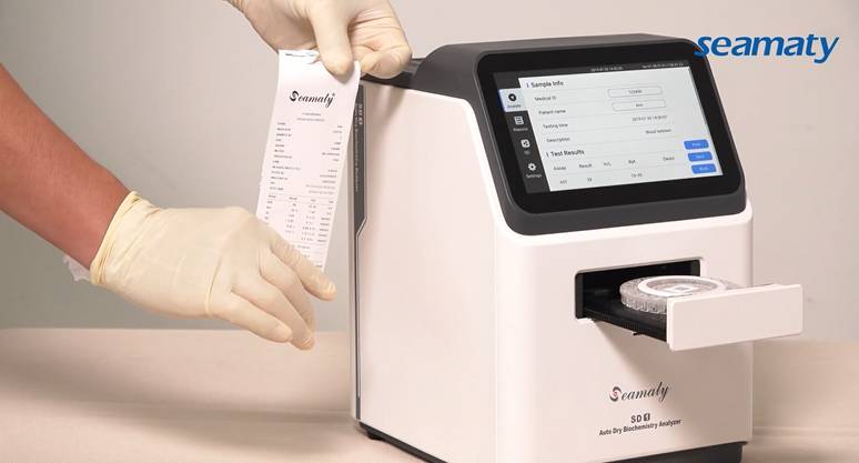
Blood tests are an essential part of our regular routine medical checkups. The up and down arrows in the test results make us wary. So which items are worthy of our attention?

1. Routine blood tests
Routine blood tests include three major systems: white blood cells, red blood cells and platelets.
The main function of white blood cells is to defend the body against invasion by foreign bacteria or viruses. It has the task of defense and surveillance. Leukocytes are divided into granulocytes, lymphocytes and monocytes. Granulocytes include neutrophils, eosinophils, and basophils.
Pathological increase in total leukocyte count can be seen in most bacterial infections, inflammation, uremia, acute bleeding and hemolysis, burns, post-surgery, infectious mononucleosis, heavy metal poisoning, and leukemia.
Pathological decrease in total leukocyte count is seen in viral infections, typhoid fever, paratyphoid fever, malaria, aplastic anemia, granulocyte deficiency, post-radiation therapy, post-chemotherapy, and non-leukocytotic leukemia.
Red blood cells play a role in transporting oxygen in the blood and also have an immune function. A decrease in the number of red blood cells suggests the development of anemia. There are various causes of anemia, including deficiency or impaired utilization of hematopoietic substances, excessive destruction of red blood cells or chronic bleeding due to acute bleeding and peptic ulcers. A hospital visit is required to further determine the cause of anemia.
Platelets have the function of stopping and clotting blood as well as repairing broken blood vessels. Thrombocytosis is often seen in iron deficiency anemia, malignant tumors, chronic granulocytic leukemia, etc. Thrombocytopenia tends to bleed more than once and is commonly seen in aplastic anemia, acute leukemia, hypersplenism, etc.
2. Blood biochemistry
2.1 Liver function
Liver function tests mainly reflect abnormal liver function by transaminases. Transaminases include glutamic aminotransferase (ALT), glutamic oxalacetic aminotransferase (AST) and glutamyl transpeptidase (GGT).
When 1% of hepatocytes become necrotic, the serum ALT level can increase 1-fold. When ALT > 10 times normal, there must be liver damage (such as chronic hepatitis B and other hepatitis).
When GGT>10 times normal, there are more possibilities of alcoholic liver, intra- and extra-hepatic cholestasis, primary liver cancer, etc. When the ALT and AST indexes are significantly elevated, it is necessary to consult the hepatology department.
2.2 Blood lipids
Elevated total cholesterol indicates an increased risk of coronary atherosclerosis. High triglycerides tend to cause "thickening of the blood". They are deposited on the walls of blood vessels and gradually form small plaques. This builds up slowly, making the inner diameter of the blood vessels thinner and blood flow poorer. At the same time, if the plaque is dislodged, it can cause myocardial infarction, cerebral infarction, pulmonary embolism and other adverse events.
A decrease in serum HDL cholesterol predicts an increased risk of coronary heart disease. However, an increase in this test facilitates the removal of cholesterol from peripheral tissues, including arterial walls, thereby preventing the development of atherosclerosis.
LDL cholesterol can block blood vessels, slow blood flow, and form dangerous blood clots. Its elevation means an increased risk of cardiovascular disease events. This is the primary goal in the management of dyslipidemia in the elderly.
2.3 Uric acid
Uric acid is the end product of purine metabolism in the body and is excreted via the kidneys. Long-term increases in uric acid can lead to gout and urinary stones and kidney damage.
3. Tumor markers
If tumor markers are abnormally elevated during screening, but there are no symptoms and signs, regular review and follow-up should be conducted to observe dynamic changes. At the same time, it can be combined with ultrasound, CT, MRI and other imaging examinations for comprehensive analysis.
It is generally believed that the reference significance of several times or even tens of times increase in tumor markers is relatively large. A slight increase may be related to diagnostic equipment, reagents, rest condition, infection and inflammation. Tumorigenesis is a complex process involving multiple genes and going through multiple stages. This may involve multiple tumor markers, and diagnosis with a single marker is difficult. Moreover, its specificity is not strong, its sensitivity is not high, and its role in early diagnosis of tumor is not obvious. Common tumor marker screening includes carcinoembryonic antigen, methemoglobin and serum CA-125.
Carcinoembryonic antigen is a broad-spectrum tumor marker. It was a specific marker for colon and rectal cancers in early stage. Later, through numerous tests, it has been shown that it is also elevated in the serum of many malignant tumors such as breast cancer and lung cancer. Therefore, carcinoembryonic antigen is not a specific marker for certain tumors.
Alpha fetoprotein is mainly associated with liver cancer. At present, it is mainly used clinically as a serum marker of primary liver cancer for diagnosis and efficacy monitoring of primary liver cancer.
Serum CA-125 is not only a specific marker for ovarian cancer, but the level of CA125 is also elevated in patients with adenocarcinoma of fallopian tube, endometrial cancer, cervical cancer, pancreatic cancer, intestinal cancer, breast cancer and lung cancer.


